

Learn the difference between gastroscopy and colonoscopy, including uses, preparation, sedation, findings and recovery in Seremban.

Hernia in Seremban: Warning Signs and Treatment Options
Learn hernia symptoms, emergency warning signs, diagnosis and the difference between open and laparoscopic hernia repair in Seremban.

Suspected Fracture in Seremban: Emergency Care or Orthopaedic Appointment?
Learn fracture warning signs, immediate first aid, when to visit emergency care and how orthopaedic follow-up supports recovery in Seremban.

Sports Injuries in Seremban: Sprain, Meniscus or ACL Tear?
Learn how sprains, strains, meniscus and ACL injuries differ, when imaging is needed and how sports injuries are treated in Seremban.
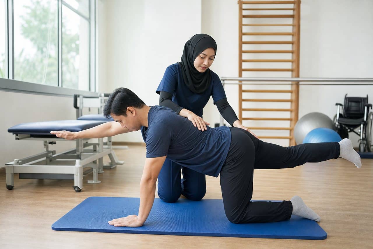
Back Pain in Seremban: Warning Signs, Diagnosis and Treatment Options
Understand back-pain causes, emergency warning signs, imaging and treatment options, and when to see a specialist in Seremban.

Knee Pain in Seremban: When Should You See an Orthopaedic Specialist?
Learn common knee-pain causes, warning signs, assessment and treatment options, and when to see an orthopaedic specialist in Seremban.
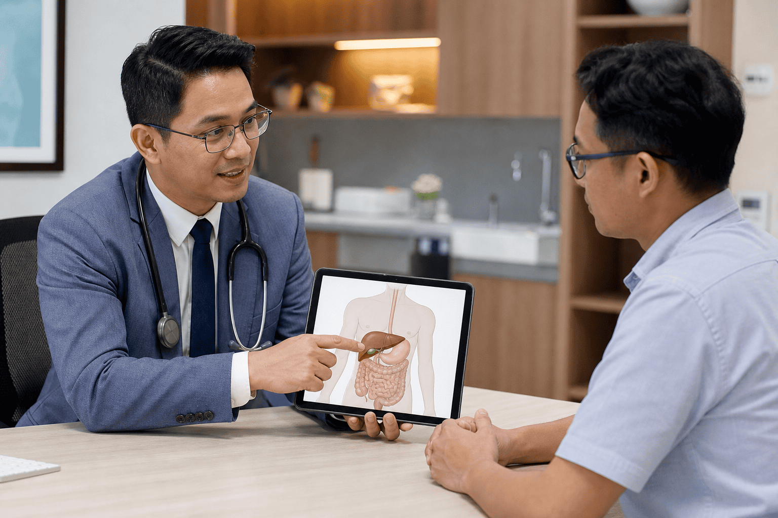
Gallstones in Seremban: Symptoms, Tests and When Surgery May Be Needed
Learn the symptoms of gallstones, urgent warning signs, diagnostic tests and when gallbladder surgery may be recommended in Seremban.
Post-Thrombotic Syndrome: Persistent Leg Swelling After a Blood Clot
Some people continue to have leg swelling, aching, heaviness or skin changes after a deep vein thrombosis (DVT). This long-term complication is called post-thrombotic syndrome. It develops when the clot and inflammation damage veins or their valves, making it harder for blood to return from the leg.
New or suddenly worsening symptoms still need assessment because a recurrent DVT can feel similar. A previous clot should never be used to dismiss a new change.
Key takeaways
- Post-thrombotic syndrome can cause chronic swelling, heaviness, discomfort and skin discolouration.
- Sudden worsening requires assessment for another clot or another cause.
- Compression is not suitable for everyone and should be individually assessed.
- Protecting the skin and maintaining suitable movement can support long-term care.
Why symptoms remain after DVT
A DVT blocks a deep vein and can injure one-way valves that normally help blood travel back to the heart. Even after anticoagulant treatment, residual obstruction or valve damage may leave higher pressure in the lower-leg veins.
Symptoms often worsen after long periods of standing or sitting. They may improve with walking or leg elevation, but patterns vary.
Symptoms to discuss with a clinician
Common features include swelling, aching, heaviness, cramps, itching, tingling, visible veins and darker skin near the ankle. Severe disease can cause eczema-like inflammation or a venous ulcer.
The clinician may compare both legs, examine the skin and pulses, review the original DVT and arrange ultrasound when a new clot or persistent obstruction is suspected.
Management is individual
A plan may include suitable walking and calf-muscle activity, weight management, periods of elevation, moisturising dry skin and properly fitted compression. Compression strength and suitability should be assessed, especially when arterial circulation may also be reduced.
Continue anticoagulants exactly as prescribed. They prevent clot extension and recurrence but do not instantly repair damaged valves. Never change treatment without medical advice.
Red flags after a previous blood clot
Seek urgent care for a sudden increase in one-sided swelling or pain. Chest pain, unexplained breathlessness, coughing blood, collapse or a very fast heartbeat may indicate pulmonary embolism and require emergency attention.
Prompt review is also important for broken skin, spreading redness, fever or a wound that is not healing.
Getting appropriate care in Seremban
For an individual assessment, contact CMH Vascular Surgery service. The appropriate specialty, test or urgency depends on your symptoms, medical history and examination.
This article provides general health information and is not a diagnosis or a substitute for personalised medical advice. Seek urgent medical care for severe or rapidly worsening symptoms.
References
Sudden Leg Pain, Pallor or Coldness: Recognising Acute Limb Ischaemia
Acute limb ischaemia means that blood flow to an arm or leg has suddenly fallen. A clot, embolus or abrupt blockage can deprive tissue of oxygen. Without rapid assessment and treatment, nerves, muscles and the limb itself may be permanently damaged.
This is not the same as gradually developing leg discomfort during walking. Sudden severe symptoms—especially a cold, pale, numb or weak limb—require emergency care.
Key takeaways
- Sudden limb pain with pallor, coldness, numbness or weakness is an emergency.
- Do not massage the limb, apply tight compression or wait for a routine clinic visit.
- Tell emergency staff when symptoms began and whether you use blood thinners.
- Fast treatment aims to restore blood flow and protect tissue.
The six warning signs
Clinicians often describe six Ps: pain, pallor, pulselessness, paraesthesia, paralysis and poikilothermia, meaning unusual coldness. A patient may not have every sign, and a pulse can be difficult to assess without equipment.
Numbness or loss of movement suggests threatened nerves and muscles. These late features make immediate assessment especially important.

Why it can happen
Possible causes include a clot forming in a narrowed artery, a clot travelling from the heart, blockage of a previous bypass or stent, injury, or separation of layers within an artery. Heart-rhythm disorders and established vascular disease can increase risk.
Only urgent examination and imaging can establish the cause. The symptoms should not be self-diagnosed as a cramp or muscle strain.
What to do immediately
Seek emergency medical help. Keep the limb in a neutral, comfortable position and avoid food or drink until medical staff advise, because an urgent procedure may be needed. Bring a medicines list and state the exact time the limb last felt normal.
Do not walk repeatedly to “test” the leg, massage it or use tight bandages. Do not take extra anticoagulant medicine unless instructed by a clinician.
How doctors assess the limb
Assessment may include comparing colour and temperature, checking movement and sensation, feeling pulses and using a handheld Doppler. CT angiography, catheter angiography or other imaging may be required to locate the blockage.
Treatment depends on how threatened the limb is and may involve anticoagulation, clot removal, catheter treatment, angioplasty, stenting or surgery.
Getting appropriate care in Seremban
For an individual assessment, contact CMH Vascular Surgery service. The appropriate specialty, test or urgency depends on your symptoms, medical history and examination.
This article provides general health information and is not a diagnosis or a substitute for personalised medical advice. Seek urgent medical care for severe or rapidly worsening symptoms.
References
Vascular Ultrasound Explained: How Doppler Scans Assess Blood Flow
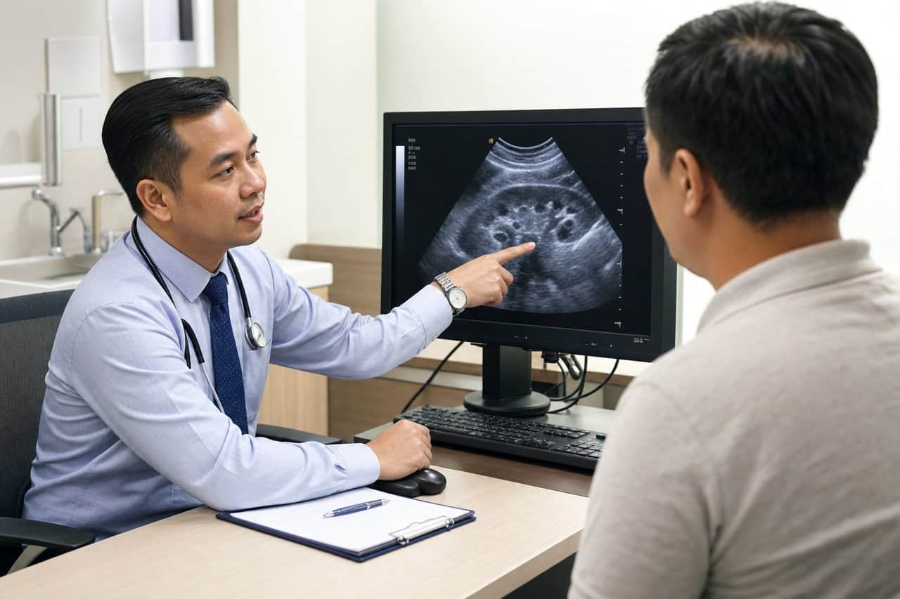
A vascular ultrasound—also called duplex or Doppler ultrasound—uses sound waves to create images of arteries or veins and assess moving blood. It can help identify narrowing, blockage, reflux or a blood clot without ionising radiation.
The scan requested depends on the clinical question. An arterial study, venous clot study and varicose-vein reflux assessment examine different structures and should not be treated as interchangeable tests.
Key takeaways
- Doppler ultrasound evaluates blood direction and speed as well as vessel appearance.
- It may be used for suspected DVT, arterial narrowing, aneurysm or venous reflux.
- Most studies are non-invasive and use gel and a handheld probe.
- A normal scan does not rule out every cause of pain or swelling.
What happens during the scan?
The sonographer exposes the area being examined, applies water-based gel and moves a probe over the skin. You may hear pulsing sounds representing blood flow. Gentle pressure or position changes may be required, especially during a vein study.
The duration varies with the body area and complexity. Some abdominal vascular studies require fasting, while many limb studies do not. Follow the instructions supplied with the appointment.
Questions vascular ultrasound can answer
A venous study can look for deep vein thrombosis. An arterial study can assess reduced blood flow and help locate narrowing. Other protocols evaluate carotid arteries, an enlarged aorta, dialysis access or the direction of flow in varicose veins.
The result must be interpreted alongside symptoms, pulse examination, blood pressure measurements and medical history.
Benefits and limitations
Ultrasound is widely used because it is non-invasive and does not use X-rays. However, deep vessels, heavy calcification, swelling or body habitus can make some areas difficult to assess. Further imaging may be recommended when ultrasound cannot answer the clinical question.
Do not assume that one scan is the best first test for every circulation concern. Clinical assessment determines the correct protocol.
When symptoms should not wait
A suddenly cold, pale, painful or numb limb is an emergency. New one-sided leg swelling with chest pain, breathlessness or coughing blood also requires urgent medical attention. Do not wait for a routine outpatient ultrasound appointment in these situations.
Getting appropriate care in Seremban
For an individual assessment, contact CMH Vascular Surgery service. The appropriate specialty, test or urgency depends on your symptoms, medical history and examination.
This article provides general health information and is not a diagnosis or a substitute for personalised medical advice. Seek urgent medical care for severe or rapidly worsening symptoms.