
Glaucoma Screening: Why Eye Pressure Alone Is Not Enough

Glaucoma Screening: Why Eye Pressure Alone Is Not Enough

Glaucoma can damage the optic nerve gradually, and some people develop it despite eye pressure readings within the usual range. Here is how patients and families can prepare for an appropriate assessment in Seremban. This guide explains the practical differences patients and families should recognise before seeking care in Seremban.

Risk is influenced by age, family history, eye anatomy, short-sightedness, steroid exposure and certain medical conditions. Many chronic cases have no early symptoms, which is why risk-based examination matters. Symptoms should be interpreted with the person’s age, medical history, medicines and the way the problem has changed over time. A focused assessment is therefore more useful than judging the condition from a single symptom or online picture.
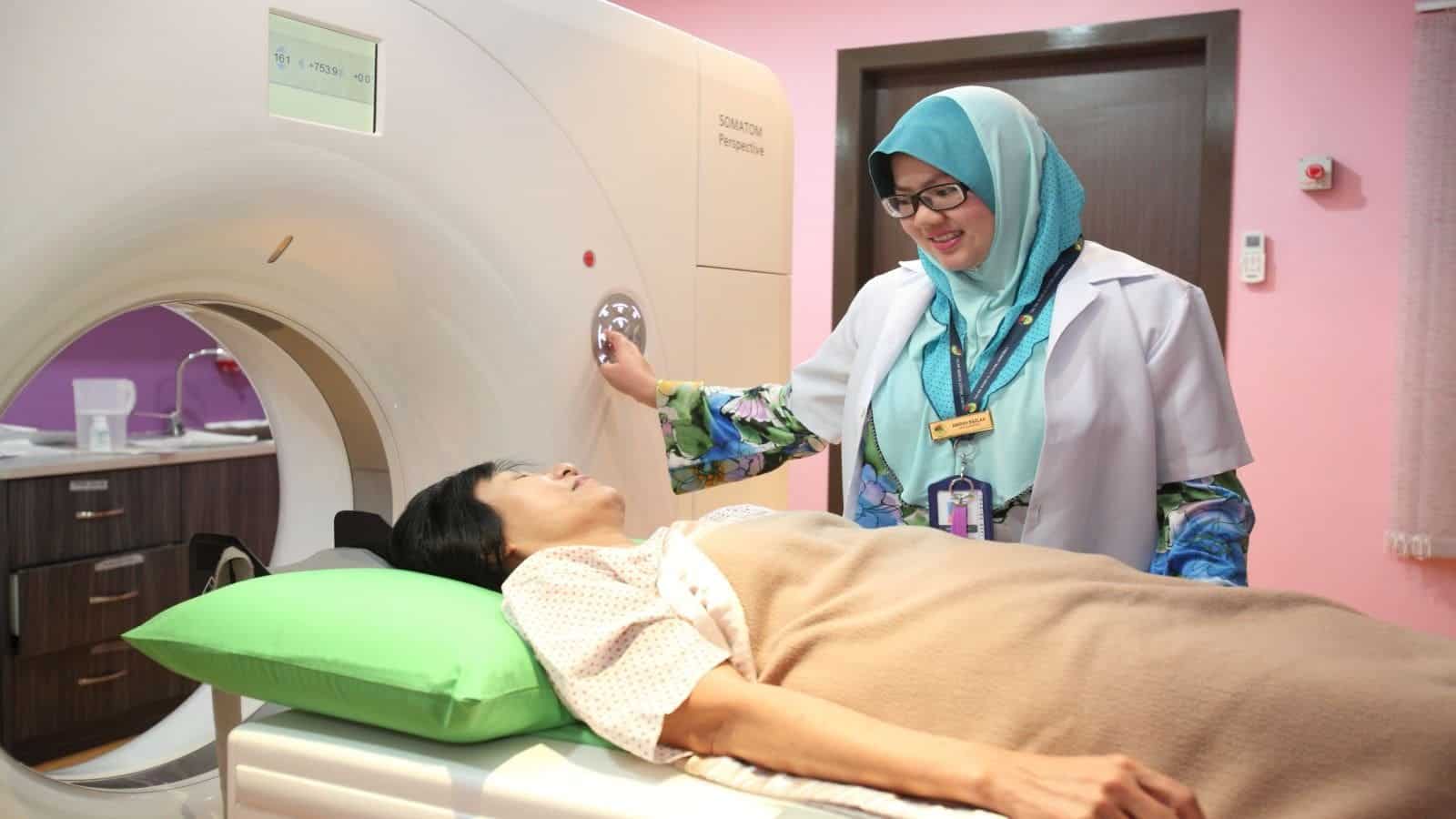
glaucoma screening Seremban — CMH CT scanner
CMH CT scanner: the first step is matching the symptom pattern with an appropriate clinical assessment.

Key Takeaways

Key pointWhat it means
PatternGlaucoma can damage the optic nerve gradually, and some people develop it despite eye pressure readings within the usual range.
Possible causesRisk is influenced by age, family history, eye anatomy, short-sightedness, steroid exposure and certain medical conditions.
AssessmentA comprehensive assessment may include pressure measurement, optic-nerve examination, visual-field testing, corneal thickness and drainage-angle evaluation.
TreatmentIf glaucoma is diagnosed, eye drops, laser treatment or surgery may be used to lower risk of further damage.
Urgent helpSevere eye pain, headache, a red eye, blurred vision, coloured halos, nausea or vomiting can signal acute angle closure and requires urgent care.

Eye pressure is one risk marker, not the diagnosis

Risk is influenced by age, family history, eye anatomy, short-sightedness, steroid exposure and certain medical conditions. Many chronic cases have no early symptoms, which is why risk-based examination matters. A short timeline noting frequency, severity and recent changes can make the consultation more useful. The timing, side of the body, triggers, associated symptoms and effect on daily function help separate common explanations from conditions that need earlier review. Guidance from NEI: Glaucoma and eye pressure supports looking at the whole clinical pattern rather than one isolated sign.

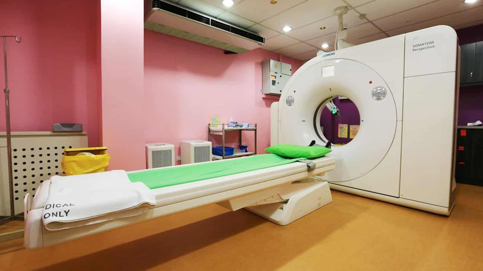
glaucoma screening Seremban — CMH diagnostic imaging area
CMH diagnostic imaging area: investigations are chosen to answer a specific clinical question.

Optic nerve and visual-field testing explained

A comprehensive assessment may include pressure measurement, optic-nerve examination, visual-field testing, corneal thickness and drainage-angle evaluation. One pressure reading cannot show the full pattern or existing nerve damage. Prepare allergies, medical conditions and earlier treatment responses so they can be reviewed accurately. A clinician may begin with simple observations and only add tests that can change the diagnosis or next step. NEI: Glaucoma provides additional patient guidance about the relevant assessment pathway.

Who may need closer glaucoma surveillance

If glaucoma is diagnosed, eye drops, laser treatment or surgery may be used to lower risk of further damage. Regular follow-up checks whether pressure and structural or field measurements remain stable. Follow-up is important when the response is incomplete or new symptoms appear. The safest plan also considers other medical conditions, pregnancy where relevant, allergies, kidney or liver function, current medicines and patient preferences. Follow-up should state what improvement is expected and when the plan needs review.

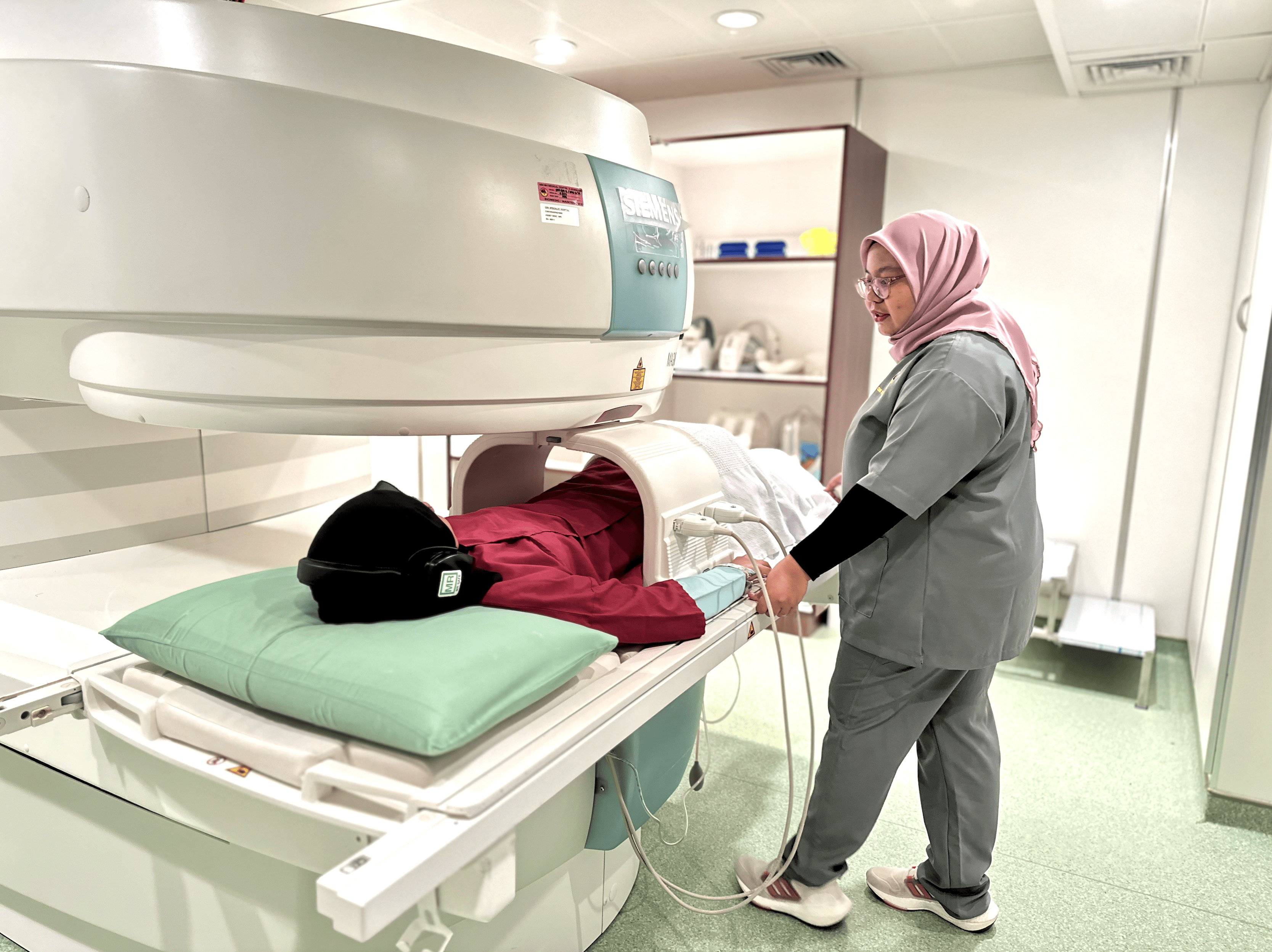
glaucoma screening Seremban — eyesight assessment
eyesight assessment: follow-up connects test findings with symptoms and response to treatment.

Preparing for an appointment about Glaucoma screening

For glaucoma screening, prepare a brief timeline: the first episode, the most recent episode, frequency, duration and any clear trigger. Note which home measures or prescribed treatments were tried, the dose and the response. Bring relevant reports and images rather than relying on memory. If symptoms come and go, a dated photograph, measurement or symptom diary may help the ophthalmology team understand what happens between visits.

When Glaucoma screening needs urgent assessment

Severe eye pain, headache, a red eye, blurred vision, coloured halos, nausea or vomiting can signal acute angle closure and requires urgent care. Do not drive yourself when faint, severely breathless or affected by sudden neurological symptoms. These warning signs do not identify one diagnosis, but they can signal that waiting is unsafe. NEI: Detection of glaucoma offers further authoritative background; local emergency instructions should take priority when symptoms are acute.

Frequently Asked Questions

Can glaucoma screening improve without specialist treatment?

Some short-lived or mild symptoms improve, but persistence, recurrence or loss of normal function changes the decision. Because glaucoma screening can have more than one cause, an examination is safer than repeatedly trying treatments without a diagnosis.

Does glaucoma screening always require a scan or procedure?

No. Tests are selected after the history and examination. A comprehensive assessment may include pressure measurement, optic-nerve examination, visual-field testing, corneal thickness and drainage-angle evaluation. A scan, laboratory test or procedure is useful only when it is likely to clarify the cause or change management.

What should I record before a ophthalmology appointment?

For glaucoma screening, write down when the problem started, how often it occurs, what makes it better or worse, and how it affects sleep, work, school or exercise. Bring previous reports, a current medicine list, allergies and relevant family history so that changes can be assessed in context.

Which warning signs make glaucoma screening urgent?

Severe eye pain, headache, a red eye, blurred vision, coloured halos, nausea or vomiting can signal acute angle closure and requires urgent care. When symptoms are severe, rapidly worsening or accompanied by collapse, breathing difficulty or a new neurological problem, use an emergency service rather than waiting for a routine appointment.

Who should assess glaucoma screening in Seremban?

The appropriate starting point for glaucoma screening depends on its pattern and urgency. CMH’s ophthalmology service can assess problems within this specialty, while emergency symptoms require immediate hospital care. If you are unsure where to start, contact the hospital and describe the main symptom and how quickly it developed.

Getting ophthalmology care in Seremban

For an individual assessment of glaucoma screening, visit the ophthalmology service at CMH Specialist Hospital. You can also contact CMH for appointment information. The clinician will decide whether observation, testing, treatment or referral is appropriate after reviewing the full history and examination.

Related CMH Articles

Conclusion

Glaucoma can damage the optic nerve gradually, and some people develop it despite eye pressure readings within the usual range. The important next step is to match the pace of care to the warning signs and to base treatment on a clinical diagnosis, not on one symptom alone. This article provides general health information and does not replace an examination, diagnosis or personalised advice from a qualified healthcare professional.
References

  1. NEI: Glaucoma and eye pressure
  2. NEI: Glaucoma
  3. NEI: Detection of glaucoma

Cataracts: Early Vision Changes and How They Are Diagnosed

Cataracts: Early Vision Changes and How They Are Diagnosed

Cataracts gradually cloud the natural lens of the eye. Early changes can be subtle and may be mistaken for an outdated glasses prescription. The sections below outline practical next steps for patients considering specialist care in Seremban. This guide explains the practical differences patients and families should recognise before seeking care in Seremban.

Typical symptoms include glare, halos, faded colours, poorer night vision and frequent prescription changes. Cataracts usually develop slowly and are often painless; sudden visual loss suggests another cause. Symptoms should be interpreted with the person’s age, medical history, medicines and the way the problem has changed over time. A focused assessment is therefore more useful than judging the condition from a single symptom or online picture.

cataract diagnosis Seremban — CMH Imaging Department
CMH Imaging Department: the first step is matching the symptom pattern with an appropriate clinical assessment.

Key Takeaways

Key pointWhat it means
PatternCataracts gradually cloud the natural lens of the eye.
Possible causesTypical symptoms include glare, halos, faded colours, poorer night vision and frequent prescription changes.
AssessmentDiagnosis uses visual-acuity testing and a slit-lamp examination after reviewing how vision affects reading, driving and daily tasks.
TreatmentUpdated glasses, better lighting and glare control may help initially.
Urgent helpPrompt assessment is required for sudden deterioration, severe pain, a red eye, flashes, a curtain-like shadow, double vision with neurological symptoms or eye injury.

Early cataract symptoms can be subtle

Typical symptoms include glare, halos, faded colours, poorer night vision and frequent prescription changes. Cataracts usually develop slowly and are often painless; sudden visual loss suggests another cause. Record when symptoms begin, their duration, triggers and any effect on sleep or daily activity. The timing, side of the body, triggers, associated symptoms and effect on daily function help separate common explanations from conditions that need earlier review. Guidance from NEI: Cataracts supports looking at the whole clinical pattern rather than one isolated sign.

cataract diagnosis Seremban — CMH MRI scanner
CMH MRI scanner: investigations are chosen to answer a specific clinical question.

How a dilated eye examination confirms cataract

Diagnosis uses visual-acuity testing and a slit-lamp examination after reviewing how vision affects reading, driving and daily tasks. The clinician also checks for glaucoma, retinal disease and other contributors to reduced sight. Previous test results and the names and doses of current medicines provide important context. A clinician may begin with simple observations and only add tests that can change the diagnosis or next step. NEI: Dilated eye exam provides additional patient guidance about the relevant assessment pathway.

When surgery becomes a practical option

Updated glasses, better lighting and glare control may help initially. Surgery is considered when cataract-related impairment meaningfully affects daily life and expected benefits outweigh individual risks. Confirm which changes are expected, which are not, and who to contact if symptoms worsen. The safest plan also considers other medical conditions, pregnancy where relevant, allergies, kidney or liver function, current medicines and patient preferences. Follow-up should state what improvement is expected and when the plan needs review.

cataract diagnosis Seremban — CMH imaging examination room
CMH imaging examination room: follow-up connects test findings with symptoms and response to treatment.

Preparing for an appointment about Cataract diagnosis

For cataract diagnosis, prepare a brief timeline: the first episode, the most recent episode, frequency, duration and any clear trigger. Note which home measures or prescribed treatments were tried, the dose and the response. Bring relevant reports and images rather than relying on memory. If symptoms come and go, a dated photograph, measurement or symptom diary may help the ophthalmology team understand what happens between visits.

When Cataract diagnosis needs urgent assessment

Prompt assessment is required for sudden deterioration, severe pain, a red eye, flashes, a curtain-like shadow, double vision with neurological symptoms or eye injury. Emergency symptoms require immediate attention even if a routine specialist visit is already arranged. These warning signs do not identify one diagnosis, but they can signal that waiting is unsafe. NEI: Cataract patient booklet offers further authoritative background; local emergency instructions should take priority when symptoms are acute.

Frequently Asked Questions

Can cataract diagnosis improve without specialist treatment?

Some short-lived or mild symptoms improve, but persistence, recurrence or loss of normal function changes the decision. Because cataract diagnosis can have more than one cause, an examination is safer than repeatedly trying treatments without a diagnosis.

Does cataract diagnosis always require a scan or procedure?

No. Tests are selected after the history and examination. Diagnosis uses visual-acuity testing and a slit-lamp examination after reviewing how vision affects reading, driving and daily tasks. A scan, laboratory test or procedure is useful only when it is likely to clarify the cause or change management.

What should I record before a ophthalmology appointment?

For cataract diagnosis, write down when the problem started, how often it occurs, what makes it better or worse, and how it affects sleep, work, school or exercise. Bring previous reports, a current medicine list, allergies and relevant family history so that changes can be assessed in context.

Which warning signs make cataract diagnosis urgent?

Prompt assessment is required for sudden deterioration, severe pain, a red eye, flashes, a curtain-like shadow, double vision with neurological symptoms or eye injury. When symptoms are severe, rapidly worsening or accompanied by collapse, breathing difficulty or a new neurological problem, use an emergency service rather than waiting for a routine appointment.

Who should assess cataract diagnosis in Seremban?

The appropriate starting point for cataract diagnosis depends on its pattern and urgency. CMH’s ophthalmology service can assess problems within this specialty, while emergency symptoms require immediate hospital care. If you are unsure where to start, contact the hospital and describe the main symptom and how quickly it developed.

Getting ophthalmology care in Seremban

For an individual assessment of cataract diagnosis, visit the ophthalmology service at CMH Specialist Hospital. You can also contact CMH for appointment information. The clinician will decide whether observation, testing, treatment or referral is appropriate after reviewing the full history and examination.

Related CMH Articles

Conclusion

Cataracts gradually cloud the natural lens of the eye. The important next step is to match the pace of care to the warning signs and to base treatment on a clinical diagnosis, not on one symptom alone. This article provides general health information and does not replace an examination, diagnosis or personalised advice from a qualified healthcare professional.
References

  1. NEI: Cataracts
  2. NEI: Dilated eye exam
  3. NEI: Cataract patient booklet

Sudden Blurred Vision: When It Is an Eye Emergency

Sudden Blurred Vision: When It Is an Eye Emergency

Sudden blurred vision may come from the eye, optic nerve, circulation or brain. The urgency depends on whether it is one eye or both and which symptoms accompany it. The aim is to help patients describe the problem clearly and recognise when faster care is needed. This guide explains the practical differences patients and families should recognise before seeking care in Seremban.

Possible causes range from corneal problems and migraine to retinal detachment, acute glaucoma, vascular blockage or stroke. A painless problem can still threaten sight, so waiting for pain is unsafe. Symptoms should be interpreted with the person’s age, medical history, medicines and the way the problem has changed over time. A focused assessment is therefore more useful than judging the condition from a single symptom or online picture.

sudden blurred vision Seremban — eyesight assessment
eyesight assessment: the first step is matching the symptom pattern with an appropriate clinical assessment.

Key Takeaways

Key pointWhat it means
PatternSudden blurred vision may come from the eye, optic nerve, circulation or brain.
Possible causesPossible causes range from corneal problems and migraine to retinal detachment, acute glaucoma, vascular blockage or stroke.
AssessmentAn eye assessment checks visual acuity, pupils, eye pressure, the front of the eye and the retina.
TreatmentTreatment is cause-specific and time-sensitive for several conditions.
Urgent helpSeek emergency care for sudden loss or marked blurring, a curtain or shadow, new flashes and many floaters, severe eye pain, halos with nausea, injury, weakness or speech change.

One eye or both eyes: why the distinction matters

Possible causes range from corneal problems and migraine to retinal detachment, acute glaucoma, vascular blockage or stroke. A painless problem can still threaten sight, so waiting for pain is unsafe. Write down the first episode, the most recent episode and anything that has clearly changed. The timing, side of the body, triggers, associated symptoms and effect on daily function help separate common explanations from conditions that need earlier review. Guidance from NEI: Retinal detachment supports looking at the whole clinical pattern rather than one isolated sign.

sudden blurred vision Seremban — CMH CT scanner
CMH CT scanner: investigations are chosen to answer a specific clinical question.

Eye, circulation and neurological warning signs

An eye assessment checks visual acuity, pupils, eye pressure, the front of the eye and the retina. Neurological examination and urgent imaging may be required when weakness, speech difficulty or other stroke features are present. Bring relevant reports, scans and a complete list of prescribed and non-prescription products. A clinician may begin with simple observations and only add tests that can change the diagnosis or next step. American Stroke Association: Stroke symptoms provides additional patient guidance about the relevant assessment pathway.

What an urgent eye assessment may include

Treatment is cause-specific and time-sensitive for several conditions. Avoid driving, cover neither eye unless instructed, and bring a medicines list and the exact time symptoms began. Ask how progress will be measured and when the diagnosis or treatment plan should be reconsidered. The safest plan also considers other medical conditions, pregnancy where relevant, allergies, kidney or liver function, current medicines and patient preferences. Follow-up should state what improvement is expected and when the plan needs review.

sudden blurred vision Seremban — CMH diagnostic imaging area
CMH diagnostic imaging area: follow-up connects test findings with symptoms and response to treatment.

Preparing for an appointment about Sudden blurred vision

For sudden blurred vision, prepare a brief timeline: the first episode, the most recent episode, frequency, duration and any clear trigger. Note which home measures or prescribed treatments were tried, the dose and the response. Bring relevant reports and images rather than relying on memory. If symptoms come and go, a dated photograph, measurement or symptom diary may help the ophthalmology team understand what happens between visits.

When Sudden blurred vision needs urgent assessment

Seek emergency care for sudden loss or marked blurring, a curtain or shadow, new flashes and many floaters, severe eye pain, halos with nausea, injury, weakness or speech change. When in doubt about a serious warning sign, urgent clinical triage is safer than self-diagnosis. These warning signs do not identify one diagnosis, but they can signal that waiting is unsafe. NEI: Eye health information offers further authoritative background; local emergency instructions should take priority when symptoms are acute.

Frequently Asked Questions

Can sudden blurred vision improve without specialist treatment?

Some short-lived or mild symptoms improve, but persistence, recurrence or loss of normal function changes the decision. Because sudden blurred vision can have more than one cause, an examination is safer than repeatedly trying treatments without a diagnosis.

Does sudden blurred vision always require a scan or procedure?

No. Tests are selected after the history and examination. An eye assessment checks visual acuity, pupils, eye pressure, the front of the eye and the retina. A scan, laboratory test or procedure is useful only when it is likely to clarify the cause or change management.

What should I record before a ophthalmology appointment?

For sudden blurred vision, write down when the problem started, how often it occurs, what makes it better or worse, and how it affects sleep, work, school or exercise. Bring previous reports, a current medicine list, allergies and relevant family history so that changes can be assessed in context.

Which warning signs make sudden blurred vision urgent?

Seek emergency care for sudden loss or marked blurring, a curtain or shadow, new flashes and many floaters, severe eye pain, halos with nausea, injury, weakness or speech change. When symptoms are severe, rapidly worsening or accompanied by collapse, breathing difficulty or a new neurological problem, use an emergency service rather than waiting for a routine appointment.

Who should assess sudden blurred vision in Seremban?

The appropriate starting point for sudden blurred vision depends on its pattern and urgency. CMH’s ophthalmology service can assess problems within this specialty, while emergency symptoms require immediate hospital care. If you are unsure where to start, contact the hospital and describe the main symptom and how quickly it developed.

Getting ophthalmology care in Seremban

For an individual assessment of sudden blurred vision, visit the ophthalmology service at CMH Specialist Hospital. You can also contact CMH for appointment information. The clinician will decide whether observation, testing, treatment or referral is appropriate after reviewing the full history and examination.

Related CMH Articles

Conclusion

Sudden blurred vision may come from the eye, optic nerve, circulation or brain. The important next step is to match the pace of care to the warning signs and to base treatment on a clinical diagnosis, not on one symptom alone. This article provides general health information and does not replace an examination, diagnosis or personalised advice from a qualified healthcare professional.

References

  1. NEI: Retinal detachment
  2. American Stroke Association: Stroke symptoms
  3. NEI: Eye health information

Living With Lupus: Monitoring Tests and Flare Warning Signs

Living With Lupus: Monitoring Tests and Flare Warning Signs

Lupus can affect skin, joints, blood cells, kidneys, lungs, heart and the nervous system, so monitoring is broader than checking joint pain alone. A focused consultation can separate common causes from problems that should not be delayed. This guide explains the practical differences patients and families should recognise before seeking care in Seremban.

Symptoms may come in flares and include fatigue, rashes, mouth ulcers, hair loss, joint pain, fever or sensitivity to sunlight. Changes in urine, swelling or blood pressure can indicate kidney involvement even before pain develops. Symptoms should be interpreted with the person’s age, medical history, medicines and the way the problem has changed over time. A focused assessment is therefore more useful than judging the condition from a single symptom or online picture.

lupus monitoring Seremban — CMH X-ray facility
CMH X-ray facility: the first step is matching the symptom pattern with an appropriate clinical assessment.

Key Takeaways

Key pointWhat it means
PatternLupus can affect skin, joints, blood cells, kidneys, lungs, heart and the nervous system, so monitoring is broader than checking joint pain alone.
Possible causesSymptoms may come in flares and include fatigue, rashes, mouth ulcers, hair loss, joint pain, fever or sensitivity to sunlight.
AssessmentFollow-up may include blood counts, kidney function, urine tests, complement levels and antibodies, selected according to the patient’s condition and treatment.
TreatmentMedication adherence, sun protection, vaccination planning and discussion before pregnancy are important parts of care.
Urgent helpUrgent assessment is needed for chest pain, severe breathlessness, seizures, confusion, new weakness, very low urine output, rapidly increasing swelling or signs of serious infection.

Recognising a possible lupus flare

Symptoms may come in flares and include fatigue, rashes, mouth ulcers, hair loss, joint pain, fever or sensitivity to sunlight. Changes in urine, swelling or blood pressure can indicate kidney involvement even before pain develops. Photographs or a dated symptom log may help when the finding comes and goes between appointments. The timing, side of the body, triggers, associated symptoms and effect on daily function help separate common explanations from conditions that need earlier review. Guidance from NIAMS: Lupus diagnosis and treatment supports looking at the whole clinical pattern rather than one isolated sign.

lupus monitoring Seremban — guided rehabilitation at CMH
guided rehabilitation at CMH: investigations are chosen to answer a specific clinical question.

Blood, urine and organ monitoring explained

Follow-up may include blood counts, kidney function, urine tests, complement levels and antibodies, selected according to the patient’s condition and treatment. Results are interpreted as trends rather than isolated numbers. A family member may help recall the history, but the patient’s privacy and preferences remain central. A clinician may begin with simple observations and only add tests that can change the diagnosis or next step. NIAMS: Lupus overview provides additional patient guidance about the relevant assessment pathway.

Planning safely around medicines and pregnancy

Medication adherence, sun protection, vaccination planning and discussion before pregnancy are important parts of care. Patients should ask how to recognise their personal flare pattern and when to contact the team. Clarify the expected timeline, possible side effects and the signs that should prompt an earlier review. The safest plan also considers other medical conditions, pregnancy where relevant, allergies, kidney or liver function, current medicines and patient preferences. Follow-up should state what improvement is expected and when the plan needs review.

lupus monitoring Seremban — reviewing an X-ray at CMH
reviewing an X-ray at CMH: follow-up connects test findings with symptoms and response to treatment.

Preparing for an appointment about Lupus monitoring

For lupus monitoring, prepare a brief timeline: the first episode, the most recent episode, frequency, duration and any clear trigger. Note which home measures or prescribed treatments were tried, the dose and the response. Bring relevant reports and images rather than relying on memory. If symptoms come and go, a dated photograph, measurement or symptom diary may help the rheumatology team understand what happens between visits.

When Lupus monitoring needs urgent assessment

Urgent assessment is needed for chest pain, severe breathlessness, seizures, confusion, new weakness, very low urine output, rapidly increasing swelling or signs of serious infection. If the pattern is clearly different from usual, seek timely medical assessment rather than waiting. These warning signs do not identify one diagnosis, but they can signal that waiting is unsafe. NIAMS: Lupus research and resources offers further authoritative background; local emergency instructions should take priority when symptoms are acute.

Frequently Asked Questions

Can lupus monitoring improve without specialist treatment?

Some short-lived or mild symptoms improve, but persistence, recurrence or loss of normal function changes the decision. Because lupus monitoring can have more than one cause, an examination is safer than repeatedly trying treatments without a diagnosis.

Does lupus monitoring always require a scan or procedure?

No. Tests are selected after the history and examination. Follow-up may include blood counts, kidney function, urine tests, complement levels and antibodies, selected according to the patient’s condition and treatment. A scan, laboratory test or procedure is useful only when it is likely to clarify the cause or change management.

What should I record before a rheumatology appointment?

For lupus monitoring, write down when the problem started, how often it occurs, what makes it better or worse, and how it affects sleep, work, school or exercise. Bring previous reports, a current medicine list, allergies and relevant family history so that changes can be assessed in context.

Which warning signs make lupus monitoring urgent?

Urgent assessment is needed for chest pain, severe breathlessness, seizures, confusion, new weakness, very low urine output, rapidly increasing swelling or signs of serious infection. When symptoms are severe, rapidly worsening or accompanied by collapse, breathing difficulty or a new neurological problem, use an emergency service rather than waiting for a routine appointment.

Who should assess lupus monitoring in Seremban?

The appropriate starting point for lupus monitoring depends on its pattern and urgency. CMH’s rheumatology service can assess problems within this specialty, while emergency symptoms require immediate hospital care. If you are unsure where to start, contact the hospital and describe the main symptom and how quickly it developed.

Getting rheumatology care in Seremban

For an individual assessment of lupus monitoring, visit the rheumatology service at CMH Specialist Hospital. You can also contact CMH for appointment information. The clinician will decide whether observation, testing, treatment or referral is appropriate after reviewing the full history and examination.

Related CMH Articles

Conclusion

Lupus can affect skin, joints, blood cells, kidneys, lungs, heart and the nervous system, so monitoring is broader than checking joint pain alone. The important next step is to match the pace of care to the warning signs and to base treatment on a clinical diagnosis, not on one symptom alone. This article provides general health information and does not replace an examination, diagnosis or personalised advice from a qualified healthcare professional.

References

  1. NIAMS: Lupus diagnosis and treatment
  2. NIAMS: Lupus overview
  3. NIAMS: Lupus research and resources

Rheumatoid Arthritis Blood Tests: RF, Anti-CCP, ESR and CRP Explained

Rheumatoid Arthritis Blood Tests: RF, Anti-CCP, ESR and CRP Explained

RF, anti-CCP, ESR and CRP can support rheumatoid arthritis assessment, but no single blood result proves or excludes the diagnosis. Understanding the pattern helps a clinician decide which examination or test is useful. This guide explains the practical differences patients and families should recognise before seeking care in Seremban.

Rheumatoid factor may be positive in people without rheumatoid arthritis, while some patients with the condition test negative. Anti-CCP is more specific, and ESR or CRP reflects inflammation but can be normal despite active joint disease. Symptoms should be interpreted with the person’s age, medical history, medicines and the way the problem has changed over time. A focused assessment is therefore more useful than judging the condition from a single symptom or online picture.

rheumatoid arthritis blood tests — CMH Physiotherapy Centre
CMH Physiotherapy Centre: the first step is matching the symptom pattern with an appropriate clinical assessment.

Key Takeaways

Key pointWhat it means
PatternRF, anti-CCP, ESR and CRP can support rheumatoid arthritis assessment, but no single blood result proves or excludes the diagnosis.
Possible causesRheumatoid factor may be positive in people without rheumatoid arthritis, while some patients with the condition test negative.
AssessmentDoctors interpret results alongside persistent swelling, morning stiffness, joint distribution and examination.
TreatmentWhen clinical suspicion is strong, referral should not be delayed only because tests are negative.
Urgent helpPrompt review is important for rapidly worsening swelling, major loss of function, fever with a hot joint, eye symptoms, chest pain or breathlessness.

What RF and anti-CCP can—and cannot—show

Rheumatoid factor may be positive in people without rheumatoid arthritis, while some patients with the condition test negative. Anti-CCP is more specific, and ESR or CRP reflects inflammation but can be normal despite active joint disease. Before the visit, note associated symptoms and whether rest, meals, movement or medicines change the pattern. The timing, side of the body, triggers, associated symptoms and effect on daily function help separate common explanations from conditions that need earlier review. Guidance from NIAMS: Rheumatoid arthritis diagnosis and treatment supports looking at the whole clinical pattern rather than one isolated sign.

rheumatoid arthritis blood tests — CMH Imaging Department
CMH Imaging Department: investigations are chosen to answer a specific clinical question.

How ESR and CRP fit into the clinical picture

Doctors interpret results alongside persistent swelling, morning stiffness, joint distribution and examination. Full blood count, kidney and liver tests may help evaluate other causes and provide a baseline before treatment. If another clinic has already investigated the problem, organise those records by date for comparison. A clinician may begin with simple observations and only add tests that can change the diagnosis or next step. NIAMS: Rheumatoid arthritis overview provides additional patient guidance about the relevant assessment pathway.

Why normal blood tests do not end the assessment

When clinical suspicion is strong, referral should not be delayed only because tests are negative. Imaging and follow-up examination can show inflammation or damage that a single blood sample cannot capture. A written plan can help patients understand medicines, monitoring and the next appointment. The safest plan also considers other medical conditions, pregnancy where relevant, allergies, kidney or liver function, current medicines and patient preferences. Follow-up should state what improvement is expected and when the plan needs review.

rheumatoid arthritis blood tests — joint examination at CMH
joint examination at CMH: follow-up connects test findings with symptoms and response to treatment.

Preparing for an appointment about Rheumatoid arthritis blood tests

For rheumatoid arthritis blood tests, prepare a brief timeline: the first episode, the most recent episode, frequency, duration and any clear trigger. Note which home measures or prescribed treatments were tried, the dose and the response. Bring relevant reports and images rather than relying on memory. If symptoms come and go, a dated photograph, measurement or symptom diary may help the rheumatology team understand what happens between visits.

When Rheumatoid arthritis blood tests needs urgent assessment

Prompt review is important for rapidly worsening swelling, major loss of function, fever with a hot joint, eye symptoms, chest pain or breathlessness. Severe or rapidly changing symptoms should be assessed through an emergency service, not a routine booking. These warning signs do not identify one diagnosis, but they can signal that waiting is unsafe. American College of Rheumatology: Rheumatoid arthritis offers further authoritative background; local emergency instructions should take priority when symptoms are acute.

Frequently Asked Questions

Can rheumatoid arthritis blood tests improve without specialist treatment?

Some short-lived or mild symptoms improve, but persistence, recurrence or loss of normal function changes the decision. Because rheumatoid arthritis blood tests can have more than one cause, an examination is safer than repeatedly trying treatments without a diagnosis.

Does rheumatoid arthritis blood tests always require a scan or procedure?

No. Tests are selected after the history and examination. Doctors interpret results alongside persistent swelling, morning stiffness, joint distribution and examination. A scan, laboratory test or procedure is useful only when it is likely to clarify the cause or change management.

What should I record before a rheumatology appointment?

For rheumatoid arthritis blood tests, write down when the problem started, how often it occurs, what makes it better or worse, and how it affects sleep, work, school or exercise. Bring previous reports, a current medicine list, allergies and relevant family history so that changes can be assessed in context.

Which warning signs make rheumatoid arthritis blood tests urgent?

Prompt review is important for rapidly worsening swelling, major loss of function, fever with a hot joint, eye symptoms, chest pain or breathlessness. When symptoms are severe, rapidly worsening or accompanied by collapse, breathing difficulty or a new neurological problem, use an emergency service rather than waiting for a routine appointment.

Who should assess rheumatoid arthritis blood tests in Seremban?

The appropriate starting point for rheumatoid arthritis blood tests depends on its pattern and urgency. CMH’s rheumatology service can assess problems within this specialty, while emergency symptoms require immediate hospital care. If you are unsure where to start, contact the hospital and describe the main symptom and how quickly it developed.

Getting rheumatology care in Seremban

For an individual assessment of rheumatoid arthritis blood tests, visit the rheumatology service at CMH Specialist Hospital. You can also contact CMH for appointment information. The clinician will decide whether observation, testing, treatment or referral is appropriate after reviewing the full history and examination.

Related CMH Articles

Conclusion

RF, anti-CCP, ESR and CRP can support rheumatoid arthritis assessment, but no single blood result proves or excludes the diagnosis. The important next step is to match the pace of care to the warning signs and to base treatment on a clinical diagnosis, not on one symptom alone. This article provides general health information and does not replace an examination, diagnosis or personalised advice from a qualified healthcare professional.

References

  1. NIAMS: Rheumatoid arthritis diagnosis and treatment
  2. NIAMS: Rheumatoid arthritis overview
  3. American College of Rheumatology: Rheumatoid arthritis

Managing Recurrent Gout Attacks: Uric Acid Targets, Medicines and Prevention

Managing Recurrent Gout Attacks: Uric Acid Targets, Medicines and Prevention

Repeated gout attacks suggest ongoing urate crystal burden, even when the joint feels normal between flares. Long-term prevention differs from short-term pain control. Here is how patients and families can prepare for an appropriate assessment in Seremban. This guide explains the practical differences patients and families should recognise before seeking care in Seremban.

Gout often affects the big toe, ankle or knee with sudden severe pain, redness and swelling. Diet, alcohol, dehydration, kidney function and medicines can influence risk, but gout is not simply caused by one meal. Symptoms should be interpreted with the person’s age, medical history, medicines and the way the problem has changed over time. A focused assessment is therefore more useful than judging the condition from a single symptom or online picture.

recurrent gout management Seremban — reviewing an X-ray at CMH
reviewing an X-ray at CMH: the first step is matching the symptom pattern with an appropriate clinical assessment.

Key Takeaways

Key pointWhat it means
PatternRepeated gout attacks suggest ongoing urate crystal burden, even when the joint feels normal between flares.
Possible causesGout often affects the big toe, ankle or knee with sudden severe pain, redness and swelling.
AssessmentDiagnosis relies on the clinical pattern and may include joint-fluid analysis, blood uric acid and imaging.
TreatmentA clinician may discuss anti-inflammatory treatment for flares and urate-lowering medicine for prevention.
Urgent helpSeek urgent assessment for fever, spreading redness, severe illness, a first hot swollen joint, immunosuppression or pain that is not following the patient’s usual gout pattern.

A flare is different from long-term urate control

Gout often affects the big toe, ankle or knee with sudden severe pain, redness and swelling. Diet, alcohol, dehydration, kidney function and medicines can influence risk, but gout is not simply caused by one meal. A short timeline noting frequency, severity and recent changes can make the consultation more useful. The timing, side of the body, triggers, associated symptoms and effect on daily function help separate common explanations from conditions that need earlier review. Guidance from NIAMS: Gout overview supports looking at the whole clinical pattern rather than one isolated sign.

recurrent gout management Seremban — CMH X-ray facility
CMH X-ray facility: investigations are chosen to answer a specific clinical question.

How gout is confirmed when the diagnosis is uncertain

Diagnosis relies on the clinical pattern and may include joint-fluid analysis, blood uric acid and imaging. Uric acid can be misleading during an acute flare, while infection must be excluded in a very hot swollen joint. Prepare allergies, medical conditions and earlier treatment responses so they can be reviewed accurately. A clinician may begin with simple observations and only add tests that can change the diagnosis or next step. NIAMS: Gout diagnosis and treatment provides additional patient guidance about the relevant assessment pathway.

Preventing crystals, attacks and joint damage

A clinician may discuss anti-inflammatory treatment for flares and urate-lowering medicine for prevention. When long-term medicine is used, adherence and repeat uric-acid checks help guide dose adjustments. Follow-up is important when the response is incomplete or new symptoms appear. The safest plan also considers other medical conditions, pregnancy where relevant, allergies, kidney or liver function, current medicines and patient preferences. Follow-up should state what improvement is expected and when the plan needs review.

recurrent gout management Seremban — guided rehabilitation at CMH
guided rehabilitation at CMH: follow-up connects test findings with symptoms and response to treatment.

Preparing for an appointment about Recurrent gout management

For recurrent gout management, prepare a brief timeline: the first episode, the most recent episode, frequency, duration and any clear trigger. Note which home measures or prescribed treatments were tried, the dose and the response. Bring relevant reports and images rather than relying on memory. If symptoms come and go, a dated photograph, measurement or symptom diary may help the rheumatology team understand what happens between visits.

When Recurrent gout management needs urgent assessment

Seek urgent assessment for fever, spreading redness, severe illness, a first hot swollen joint, immunosuppression or pain that is not following the patient’s usual gout pattern. Do not drive yourself when faint, severely breathless or affected by sudden neurological symptoms. These warning signs do not identify one diagnosis, but they can signal that waiting is unsafe. American College of Rheumatology: Gout offers further authoritative background; local emergency instructions should take priority when symptoms are acute.

Frequently Asked Questions

Can recurrent gout management improve without specialist treatment?

Some short-lived or mild symptoms improve, but persistence, recurrence or loss of normal function changes the decision. Because recurrent gout management can have more than one cause, an examination is safer than repeatedly trying treatments without a diagnosis.

Does recurrent gout management always require a scan or procedure?

No. Tests are selected after the history and examination. Diagnosis relies on the clinical pattern and may include joint-fluid analysis, blood uric acid and imaging. A scan, laboratory test or procedure is useful only when it is likely to clarify the cause or change management.

What should I record before a rheumatology appointment?

For recurrent gout management, write down when the problem started, how often it occurs, what makes it better or worse, and how it affects sleep, work, school or exercise. Bring previous reports, a current medicine list, allergies and relevant family history so that changes can be assessed in context.

Which warning signs make recurrent gout management urgent?

Seek urgent assessment for fever, spreading redness, severe illness, a first hot swollen joint, immunosuppression or pain that is not following the patient’s usual gout pattern. When symptoms are severe, rapidly worsening or accompanied by collapse, breathing difficulty or a new neurological problem, use an emergency service rather than waiting for a routine appointment.

Who should assess recurrent gout management in Seremban?

The appropriate starting point for recurrent gout management depends on its pattern and urgency. CMH’s rheumatology service can assess problems within this specialty, while emergency symptoms require immediate hospital care. If you are unsure where to start, contact the hospital and describe the main symptom and how quickly it developed.

Getting rheumatology care in Seremban

For an individual assessment of recurrent gout management, visit the rheumatology service at CMH Specialist Hospital. You can also contact CMH for appointment information. The clinician will decide whether observation, testing, treatment or referral is appropriate after reviewing the full history and examination.

Related CMH Articles

Conclusion

Repeated gout attacks suggest ongoing urate crystal burden, even when the joint feels normal between flares. The important next step is to match the pace of care to the warning signs and to base treatment on a clinical diagnosis, not on one symptom alone. This article provides general health information and does not replace an examination, diagnosis or personalised advice from a qualified healthcare professional.

References

  1. NIAMS: Gout overview
  2. NIAMS: Gout diagnosis and treatment
  3. American College of Rheumatology: Gout

Morning Joint Stiffness: When It May Be Inflammatory Arthritis

Morning Joint Stiffness: When It May Be Inflammatory Arthritis

Morning stiffness that lasts well beyond getting out of bed may point to inflammation, especially when joints are swollen, warm or affected on both sides. The sections below outline practical next steps for patients considering specialist care in Seremban. This guide explains the practical differences patients and families should recognise before seeking care in Seremban.

Inflammatory arthritis commonly improves somewhat with movement and worsens after rest. Osteoarthritis can also feel stiff, but its pattern, duration and joint distribution often differ. Infection and crystal arthritis must be considered when one joint becomes acutely hot. Symptoms should be interpreted with the person’s age, medical history, medicines and the way the problem has changed over time. A focused assessment is therefore more useful than judging the condition from a single symptom or online picture.

morning joint stiffness Seremban — joint examination at CMH
joint examination at CMH: the first step is matching the symptom pattern with an appropriate clinical assessment.

Key Takeaways

Key pointWhat it means
PatternMorning stiffness that lasts well beyond getting out of bed may point to inflammation, especially when joints are swollen, warm or affected on both sides.
Possible causesInflammatory arthritis commonly improves somewhat with movement and worsens after rest.
AssessmentA rheumatology assessment maps which joints are involved and checks swelling, movement, skin, nails and systemic symptoms.
TreatmentEarly diagnosis matters because disease-modifying treatment can protect joints in conditions such as rheumatoid arthritis.
Urgent helpUrgent care is needed for a suddenly hot swollen joint with fever, inability to bear weight, severe eye pain or redness, chest pain, marked weakness or rapidly progressive symptoms.

Inflammatory stiffness versus mechanical stiffness

Inflammatory arthritis commonly improves somewhat with movement and worsens after rest. Osteoarthritis can also feel stiff, but its pattern, duration and joint distribution often differ. Infection and crystal arthritis must be considered when one joint becomes acutely hot. Record when symptoms begin, their duration, triggers and any effect on sleep or daily activity. The timing, side of the body, triggers, associated symptoms and effect on daily function help separate common explanations from conditions that need earlier review. Guidance from NIAMS: Rheumatoid arthritis overview supports looking at the whole clinical pattern rather than one isolated sign.

morning joint stiffness Seremban — CMH Physiotherapy Centre
CMH Physiotherapy Centre: investigations are chosen to answer a specific clinical question.

Joint patterns that raise suspicion for arthritis

A rheumatology assessment maps which joints are involved and checks swelling, movement, skin, nails and systemic symptoms. Blood tests and imaging support the clinical picture; normal inflammatory markers do not rule out every inflammatory condition. Previous test results and the names and doses of current medicines provide important context. A clinician may begin with simple observations and only add tests that can change the diagnosis or next step. NIAMS: Rheumatoid arthritis diagnosis and treatment provides additional patient guidance about the relevant assessment pathway.

Why early assessment can protect function

Early diagnosis matters because disease-modifying treatment can protect joints in conditions such as rheumatoid arthritis. Exercise and pain management are tailored to the diagnosis rather than prescribed from symptoms alone. Confirm which changes are expected, which are not, and who to contact if symptoms worsen. The safest plan also considers other medical conditions, pregnancy where relevant, allergies, kidney or liver function, current medicines and patient preferences. Follow-up should state what improvement is expected and when the plan needs review.

morning joint stiffness Seremban — CMH Imaging Department
CMH Imaging Department: follow-up connects test findings with symptoms and response to treatment.

Preparing for an appointment about Morning joint stiffness

For morning joint stiffness, prepare a brief timeline: the first episode, the most recent episode, frequency, duration and any clear trigger. Note which home measures or prescribed treatments were tried, the dose and the response. Bring relevant reports and images rather than relying on memory. If symptoms come and go, a dated photograph, measurement or symptom diary may help the rheumatology team understand what happens between visits.

When Morning joint stiffness needs urgent assessment

Urgent care is needed for a suddenly hot swollen joint with fever, inability to bear weight, severe eye pain or redness, chest pain, marked weakness or rapidly progressive symptoms. Emergency symptoms require immediate attention even if a routine specialist visit is already arranged. These warning signs do not identify one diagnosis, but they can signal that waiting is unsafe. NIAMS: Arthritis diagnosis and treatment offers further authoritative background; local emergency instructions should take priority when symptoms are acute.

Frequently Asked Questions

Can morning joint stiffness improve without specialist treatment?

Some short-lived or mild symptoms improve, but persistence, recurrence or loss of normal function changes the decision. Because morning joint stiffness can have more than one cause, an examination is safer than repeatedly trying treatments without a diagnosis.

Does morning joint stiffness always require a scan or procedure?

No. Tests are selected after the history and examination. A rheumatology assessment maps which joints are involved and checks swelling, movement, skin, nails and systemic symptoms. A scan, laboratory test or procedure is useful only when it is likely to clarify the cause or change management.

What should I record before a rheumatology appointment?

For morning joint stiffness, write down when the problem started, how often it occurs, what makes it better or worse, and how it affects sleep, work, school or exercise. Bring previous reports, a current medicine list, allergies and relevant family history so that changes can be assessed in context.

Which warning signs make morning joint stiffness urgent?

Urgent care is needed for a suddenly hot swollen joint with fever, inability to bear weight, severe eye pain or redness, chest pain, marked weakness or rapidly progressive symptoms. When symptoms are severe, rapidly worsening or accompanied by collapse, breathing difficulty or a new neurological problem, use an emergency service rather than waiting for a routine appointment.

Who should assess morning joint stiffness in Seremban?

The appropriate starting point for morning joint stiffness depends on its pattern and urgency. CMH’s rheumatology service can assess problems within this specialty, while emergency symptoms require immediate hospital care. If you are unsure where to start, contact the hospital and describe the main symptom and how quickly it developed.

Getting rheumatology care in Seremban

For an individual assessment of morning joint stiffness, visit the rheumatology service at CMH Specialist Hospital. You can also contact CMH for appointment information. The clinician will decide whether observation, testing, treatment or referral is appropriate after reviewing the full history and examination.

Related CMH Articles

Conclusion

Morning stiffness that lasts well beyond getting out of bed may point to inflammation, especially when joints are swollen, warm or affected on both sides. The important next step is to match the pace of care to the warning signs and to base treatment on a clinical diagnosis, not on one symptom alone. This article provides general health information and does not replace an examination, diagnosis or personalised advice from a qualified healthcare professional.

References

  1. NIAMS: Rheumatoid arthritis overview
  2. NIAMS: Rheumatoid arthritis diagnosis and treatment
  3. NIAMS: Arthritis diagnosis and treatment